Ultrafast CT:
Visualizing the arteries that cause heart attacks
|
Introduction |
Coronary atherosclerosis (blockages of
the coronary arteries) is the major cause of death in this
society, and often exists in people at fairly “low risk” and with no
symptoms. Heart attacks and
sudden death may occur without warning.
Finding blocked heart arteries is a challenge, however.
They are relatively small structures, and like the heart itself,
are in constant motion. They
are not seen using regular x-rays nor ultrasound techniques. Indeed, even advanced techniques such as “regular” CT
scanning and MRI (Magnetic Resonance Imaging) cannot visualize them
secondary to their movement. Physicians
therefore use techniques that predict the presence of atherosclerosis that
are less precise than actually visualizing the vessels.
Physicians often assess “Risk Factors” (such as smoking, high
cholesterol, diabetes, age, sex, and family history of heart disease).
Indeed, people with much higher risk than the rest of the
population can be identified, but many cases can be missed.
Indeed, most cases of coronary disease develop in people with
average risk factor profiles.
Furthermore, some people with “high risk” may never develop
coronary disease. Techniques such as stress tests are useful, but only for discovering blockages that have grown to the point where they cause a lack of blood supply to the heart. Even advanced blockages may be missed. Stress tests are incapable of finding early atherosclerosis since they rely on the presence of advanced blockages. This is particularly frustrating since such early blockages can have the most done to prevent progressive problems. Angiograms are extremely accurate at finding plaques that have grown from the walls of the arteries and block the flow of blood. These tests are expensive, time-consuming, and are associated with at least modest risk. Again, the mildest blockages are missed. All of these techniques are limited in their ability to follow the course of the process over time. Ultrafast CT is a quick, easy, and relatively inexpensive technique which allows the arteries to the heart to be directly visualized. It images calcium in the vessel wall as well as the arterial channel to some degree. It can overcome some of the deficiencies of the other techniques. This allows for simple, early detection of atherosclerosis and the potential to follow the course of the process over time. Ultrafast CT is not without its detractors however. Many physicians, including myself, have approached this test with a great deal of suspicion. The test had been hyped by many in a position to further their financial health, and the potential for inappropriate use has been realized on many occasions. However, much has been learned – it is clear that there is much to be gained from its proper use. While improper utilization can lead to inaccurate diagnosis, further diagnostic testing, and unnecessary treatment, many people can benefit. Careful selection of patients, and careful interpretation of the results in light of multiple factors in the patient’s history is essential to maximizing the potential of the technique. To learn more, continue with the more
detailed discussion. The
following subjects are covered: The challenge of finding heart
disease before it strikes |
|
Despite the great advances in heart
disease diagnosis and management over the last two decades, one of the
great challenges remains: Finding
heart disease BEFORE it strikes. |
|
Physicians
have realized the potential for these catastrophes, and indeed work hard
to try and identify those at high risk.
This explains why we measure and treat high blood pressure, high
cholesterol, and diabetes, and why we stress the absolute need to
discontinue smoking. Treating people with these “risk factors” can indeed be
effective. Some people
with heart disease however have no risk factors, or only minimal problems.
Indeed, most heart attacks that occur will occur in people with
cholesterol levels in the “normal” range, and risk factor profiles
that are not too impressive.
|
|
There
are several varieties of “stress tests” which can be utilized to try
and find people who may have blockages.
These can show whether there are blockages of sufficient severity
to decrease the amount of blood traveling to the heart when the heart
muscle is working hard.
These
tests are useful, but have several shortcomings:
Heart catheterization is a test where “dye” (actually a material that provides contrast on the inside of the arteries so that the channel where blood flows can be seen) is used to see if there is any narrowing. It too is a very useful tool for finding blockages, and remains the best way to judge the severity of the narrowings. However, it also has problems when it comes to the task of finding heart disease early:
|
|
|
|
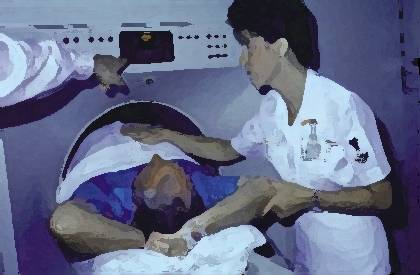
Ultrafast
CT images all parts of the heart including the muscle and valves.
Some very useful images can be made of blood flow, and heart muscle
function can be analyzed. Ultrafast
CT can also be utilized to diagnose problems such as pulmonary embolism or
dissection of the aorta.
The most exciting part about Ultrafast CT however is the ability to see the coronary arteries, the vessels that supply blood to the heart . It is important to realize that these studies are not the same as an angiogram, where contrast material (“dye”) is injected directly into the artery by catheters. Indeed, catheters make much more accurate pictures of the inside of the arteries. These blockages may also be seen with Ultrafast CT, simply not as well. Ultrafast CT shines however in discovering the process of atherosclerosis at a more basic level: It sees the development of lesions before they begin to interrupt the path of blood flow by imaging the changes that occur in the vessel wall itself. It owes its success to its ability to image calcium in the vessel walls. While calcium is not the major problem when it comes to blocked arteries (the most important substance is cholesterol), the appearance of calcium in the wall of the artery is associated with active atherosclerosis in many cases. Indeed, removing the calcium from blocked vessels, as is the basis of “chelation” therapy (see link) has really very little to do with changing the course of atherosclerosis. Calcium is however an excellent substance to use for imaging. |
|
The
amount of calcium that is deposited in atherosclerotic plaque varies from
person to person, from males to females, and certainly varies with a
person’s age. Imaging
calcium is exciting, but it also has its own limitations, and the test
must be interpreted carefully, taking many factors into consideration
before deciding that the test reliably indicates the presence or absence
of disease (see “Who should have the test?”).
|
|
|
|
Well,
as usual, it depends. Age,
gender, and the presence of other risk factors all play a role in this
recommendation. The most
important points however relate to age and gender.
It
is very unusual for people, particularly women, to have calcium in their
vessel walls under the age of 40. This
is true even if there are significant blockages present.
The process simply has not been going on long enough to be
associated with the deposition of significant calcium.
Likewise, calcium is present in many
people over the age of 65.
There is a large group “in the middle” of these age ranges for whom the test has demonstrated a substantial correlation with future heart attacks, bypass surgery, and prognosis. Indeed, this is the group in whom coronary disease is likely to strike prematurely. Those with multiple risk factors or a family history of coronary disease may be productively screened at somewhat younger ages. At least two studies have shown the promise of assessing the success of medical treatment of high blood pressure and cholesterol levels with this technique, suggesting that adequate medical therapy slows the progression of calcium deposition. If such research continues to be positive, this would be exceedingly useful. |
|
Many
insurance companies do cover this procedure now, and Medicare does have a
code that can be used for billing. However,
as in all such matters, the rules are complex and ever changing.
You or the facility that bills you will need to discuss this with
your carrier. The test is
relatively inexpensive however, and some people prefer to pay with cash
since in that case the insurance company has no legal access to the
results.
|
|
|
My Ultrafast CT came out fine.
That shows I can continue smoking and/or eating a bad diet, etc. |
No,
No, No! That’s not the
point at all. If you play
with fire, you will get burned!
Smoking is a health risk that certainly is associated with heart disease, but also causes cancers at multiple sites in the body, not to mention emphysema and other lung disease. You absolutely need to stop smoking regardless of the results of the Ultrafast CT (see “Stopping Smoking” ). This test is designed to allow you and your physician to assess your risk of heart disease and implement those health practices and therapies that will allow you to pursue a long and healthy life. It is not designed to allow you to follow improper health habits! A scan that is “normal” or “low risk” is not a guarantee of either, now or in the future. Proper diet and exercise are still among the very most important ways to live longer, and enjoy doing it! |
|
|
This has
been a fairly complex subject. Now
that we’ve covered it all in detail, let’s look at it briefly again.
|
©COPY;1997 HeartPoint Updated December 1999

![]()
| Commentary |
Food You Will Love | HeartPoint
Gallery | In The News | Health Tips | What's New
|
| Information Center | Home
|
This site presents material for your information, education and entertainment. We can assume no liability for inaccuracies, errors, or omissions. Above all, material on this site should not take the place of the care you receive from a personal physician. It is simply designed to help in the understanding of the heart and heart disease, and not as a diagnostic or therapeutic aid. You should seek prompt medical care for any specific health issues. Please feel free to browse the site and download material for personal and non-commercial use. You may not however distribute, modify, transmit or reuse any of these materials for public or commercial use. You should assume that all contents of the site are copyrighted. ©COPY;1997 HeartPoint


 Thus,
the reliability of the test is less in older age groups, particularly in
males.
Thus,
the reliability of the test is less in older age groups, particularly in
males.