Valvular Heart Disease
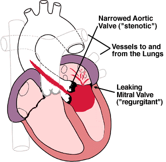
What happens with valves that leak (regurgitation)?
What happens with valves that don't open well (stenosis)?
How can you tell if I have an abnormal valve?
What is a heart "murmur"?
What causes diseases of the heart valves?
What type of therapy is available?
What kinds of medications are available for valve disease?
What kinds of surgery are available for problems with the Mitral Valve?
What kinds of surgery are available for problems with the Aortic Valve?
What kinds of artificial valves are there?
What is the best kind of artificial valve?
Do I need to take blood thinners?
What is "Endocarditis"?
What happens with valves that leak (regurgitation)?
Valves that leak cause the heart to have to pump the same blood twice . . . that is, a part of the work of the heart simply is for no good purpose, as the blood comes right back into the chamber. The heart has a number of ways it can compensate for this extra work. The first tendency is for the chambers to enlarge, since there is more blood to pump (the blood it usually would need to pump to the rest of the body plus the regurgitated blood). This is usually quite effective if the leakage is mild or moderate, and the person may not even realize there is any problem at all. In more severe cases, the heart muscle may begin to wear down. Congestive heart failure may then result, characterized by shortness of breath, swelling of the ankles, and other symptoms. The enlarged chambers may also lead to problems with arrhythmias.
What happens with valves that don't open well (stenosis)?
stenosis cause the chamber behind the narrowed valve to have to work harder. Just as when a hose is kinked, the pressure in that chamber will increase. The heart can usually compensate for this when the narrowing is mild by becoming thicker and stronger. If the narrowing progresses however, this mechanism will no longer be effective, and congestive heart failure and arrhythmias may occur.
How can you tell if I have an abnormal valve?
Most cases of valvular heart disease can be detected with a stethoscope. The abnormal blood flow often produces a sound called a heart "murmur". Not all murmurs are abnormal, nor do they necessarily indicate heart disease, but the trained ear can often tell a lot about valvular problems just with this simple device. For more about murmurs, read the section below.
The electrocardiogram is simply a representation of the electrical activity of the heart, and may show nothing at all about valvular problems, particularly in early or minor stages.
The chest x-ray, a relatively unsophisticated tool, can provide valuable information about the heart chambers and whether there is any evidence of congestive heart failure or other conditions.
An echocardiogram is a very valuable tool for evaluation of valvular heart disease. This is a non-invasive means of visualizing the heart muscle and valves using sound waves, or sonar. The valves can usually be visualized quite well, and the degree of leakage or stenosis can be estimated, in many cases with a high degree of accuracy.
Cardiac catheterization is sometimes needed to fully evaluate the heart valves, heart muscle, and heart arteries in some circumstances. Using this more "invasive" procedure, highly accurate information can be obtained.
What is a heart "murmur"?
A heart murmur is NOT a disease . . . in fact, it may be present in a person with a normal heart. A murmur is simply a sound created by blood flow. Flow that is not obstructed, like in a wide-open hose, is hard to hear. However, when a hose is kinked, flow is no longer straight and uninterrupted. This disordered flow creates sound. In the human body, a similar situation can occur, and when the disordered flow is heard with a stethoscope, it is termed a murmur.
Murmurs can be caused by having a narrowed structure in the stream of blood, such as a stenotic valve. Likewise, the backward flow through a regurgitant valve occurs through a relatively small hole and causes sounds as well. The speed of flow is also increased in conditions such as exercise or fever, and this may cause straight or linear flow to become disrupted, even in normal structures. Thus, murmurs are common in this circumstance. Infants and children often have such sounds heard by medical professionals. When it is not caused by any abnormality of the heart, it is termed an "innocent murmur". The parent is generally told "not to worry about it" and that the "child will grow out of it". Both statements are correct when there is no underlying heart disease.
What causes diseases of the heart valves?
Among the major causes of valve problems are:
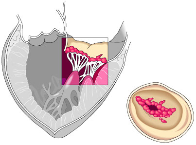
Congenital heart disease. Problems with the heart valves may be present from birth. For example, the aortic valve may be almost entirely closed from a very early age. Much more common than this however, are mild abnormalities of the valve, such as being of made of two leaflets instead of three. This so-called "bicuspid aortic valve" is often associated with an accelerated incidence of aortic stenosis that may occur when the patient is in his or her fifties.
Rheumatic heart disease. Some infections with the bacteria called "streptococcus", particularly "strep throat" are followed in several weeks to months by a delayed inflammatory reaction called "rheumatic fever". The delicate valvular structures can be damaged at that time and progressive malformation of the valve can ensue over the next several decades. Valve replacement may be necessary in these later stages. Due to the discovery and use of antibiotics, rheumatic fever is far less common than in the past, particularly in developed countries. Proper treatment of strep infections can prevent almost all of the cases of rheumatic heart disease.
Specific damage from a heart attack. Specific parts of the heart muscle concerned with proper functioning of the valves can be injured in the course of a heart attack. If there is a tear of part or all of one of the "papillary muscles", severe mitral regurgitation can occur rapidly and require emergency therapy, perhaps including surgery.
Weakening of the supporting structures of the heart. In unusual cases, there may be a tear in one of the parts of the mitral valve which attach the valve to its papillary muscle. A rupture of some of the "chordae tendiniae" can cause substantial leakage through the mitral valve. This may begin and progress slowly, or be quite severe at the onset and require emergency surgery. Weakening of the walls of the aorta can occur, which leads to gradual dilation of the aorta, which can then lead to substantial leakage through the aortic valve.
Weakening of the heart muscle. When the heart muscle weakens, regardless of cause, it will tend to lengthen. As the chamber enlarges, so too do the holes which the Mitral and Tricuspid valves are designed to cover. At some point, while the valve itself is not diseased, it simply cannot cover this area, and valvular regurgitation will begin to occur. This often leads to yet further dilation and enlargement of the ventricles, and a "vicious cycle" begins to occur. When this is the cause of the leakage, surgery on the valve alone is often not helpful, since the primary problem was the diseased heart muscle in the first place.
Infections. Infection of a heart valve is termed "endocarditis". This is not a common problem, but it can cause rapid progression of valvular disease, generally regurgitation, over a matter of days to weeks. This infection requires prompt diagnosis and treatment, but may still land up requiring valve surgery even when caught early.
Other causes. There are other even less common causes of valve disease (are you surprised?) which are not discussed here.
What type of therapy is available?
There are many, many cases of valvular heart disease which do very well without any medical intervention at all throughout a patient's life. On the other hand of course, are the numerous cases that benefit greatly from some type of medications or surgery. Surgery involves either repair and/or replacement of valves, and therefore entails open heart surgery, which may be from a standard approach or a "mini procedure". Some patients with mitral stenosis or aortic stenosis may benefit from dilation by a balloon across the valve as well.
What kinds of medications are available for valve disease?
Medications do not work to correct the defect on the valve itself, but rather minimize its consequences or treat its complications. Medications are often quite effective in some cases of valvular regurgitation for the reasons explained below under "afterload reducing agents". Valves that are stenotic however generally require surgery or balloons if their effects are to be corrected. Medications simply cannot make up for the mechanical problems that come from the narrowed valve.
Afterload reducing agents. These allow for more effective forward flow to occur despite a leaky valve -- they are not nearly as effective, or may not be effective at all, for a stenotic valve. Imagine a pump with a valve pushing water out of a hose in a pulsatile fashion. Imagine then that there was a leak in that valve. Now imagine someone went and crimped the hose -- what would happen? There would be more leakage backwards since the pressure would be higher in the hose. Afterload reducing agents decrease this backward pressure on the hose, and therefore more forward flow will occur with less work by the pump.
Diuretics. Diuretics may be prescribed if there is a tendency to "hold on to fluid" or if the patient has experienced congestive heart failure.
Digitalis. Digitalis increases the force of the heart muscle's contraction, and can be useful if the person has experienced congestive heart failure. Digitalis can also be useful for treatment of some arrhythmias, particularly atrial fibrillation, which can occur in valvular heart disease.
Blood pressure medications. Control of high blood pressure can be particularly important in people with valvular heart disease. This can decrease the amount of blood that leaks through regurgitant valves, and decrease the strain on chambers trying to pump through stenotic valves.
Blood thinners. Blood thinners, such as coumadin, aspirin, ticlodipine (Ticlid), clopidogil (Plavix), or dipyridamole (Persantine) may be prescribed. There may be a tendency to suffer from blood clots, resulting in strokes or other problems in some cases of valvular heart disease. This is more common in diseases of the mitral valve, particularly mitral stenosis. It is also an important consideration in persons who have atrial fibrillation, or who already have artificial valves.
Beta-blockers and anti-arrhythmics. Beta-blockers can be used for a variety of conditions. In the context of valvular heart disease, they are most often given for high blood pressure or irregular heart rhythms. Other types of antiarrhythmic drugs may also be prescribed.
What kinds of surgery are available for problems with the Mitral Valve?
There are several options, depending on the exact type of problem, patient, and severity of the process.
Mitral valve repair. In many cases of mitral regurgitation, the problems that cause leakage can be repaired by a skilled surgeon without the need for replacement. Often, a valve "ring" is placed in the orifice that the mitral valve covers to "tighten up" the size of this area and allow the valve to cover it more effectively. The size and shape of the leaflets can be carefully remodeled, and torn structures sewn back together. Repair offers the advantage of improved left ventricular muscle performance, since the muscle (papillary muscles) and supporting structures (chordae tendiniae) are left intact. The long term results of repairing and saving the person's original "native" valve is often better than repair, with improved heart muscle function and decreased need for repeated surgical procedures. Furthermore, after repair there may be no need for long term anticoagulation. It is important to discuss this with your surgeon if you are planning to have surgery for mitral regurgitation.
Mitral commissurotomy. During this procedure, which is done for some people with mitral stenosis, the leaflets which have fused together at their "commissures" (points of touching) are separated by the surgeon. Like mitral valve repair for regurgitation, not all cases are suitable candidates for this approach, as sometimes the mitral valve is too calcified, or the leaflets cannot be separated in a satisfactory manner. Nowadays, many candidates for commissurotomy are treated with balloon mitral valvuloplasty.
Mitral valve replacement. Some mitral valves simply need to be replaced. This may not be known until the actual time of surgery, when a repair for regurgitation or a commisurotomy for stenosis can then be seen to be impossible or ill-advised. The valvular structure is cut out, and as much of the supporting structure left as feasible. The new valve may be mechanical or bioprosthetic (see explanation and picture below), and may be done from a standard approach across the breastbone ("median sternotomy"), or with some of the new "mini" approaches.
Balloon mitral valvuloplasty. This procedure is done in a manner similar to balloon coronary artery angioplasty. That is, access is gained to the circulation from the vessels in the legs, and a catheters with deflated balloons are advanced through the vessels to a position across the mitral valve. The balloons are then inflated, creating a somewhat uncontrolled but effective commissurotomy as described above. This all obviously occurs without having to enter the chest surgically, and is much easier on the patient in most cases. Again, not all people are candidates for balloon angioplasty, particularly those with heavily calcified valves. However, it is gaining increasing popularity in many centers.
Myocardial reduction procedure with mitral valve replacement (Battista procedure). In some cases of cardiomyopathy and congestive heart failure, the heart's natural tendency to dilate becomes massive, and becomes a problem of itself. It is often associated with substantial leakage through the mitral valve, as its annulus dilates. Battista, a surgeon in South America, pioneered a surgery which actually cuts out a very substantial part of the heart muscle, restoring it to a more efficient size. The mitral valve is also often removed in the course of this surgery. Although it is counterintuitive to cut out muscle from a weak organ, initial experience has been favorable.
What kinds of surgery are available for problems with the Aortic Valve?
Aortic valve replacement. Unlike the mitral valve, repair of the aortic valve can only rarely be accomplished. If surgery is required, then replacement with either a bioprosthetic or mechanical valve is necessary (see explanation and picture below). This may be accomplished from either the classic approach through the breastbone ("median sternotomy") or newer "mini" procedures.
The Ross procedure. The Ross procedure utilizes the patient's own pulmonic valve for replacement into the aortic position.
Balloon aortic valvuloplasty. Balloon valvuloplasty of the aortic valve is an attractive technique for aortic stenosis since a large number of people with this problem are quite elderly and thus not ideal candidates for surgery. However, these same patients also typically have a heavily calcified aortic valve, and strokes secondary to loosening of this material can occur. Unfortunately, there is also a higher recurrence rate, with up to 80% of patients requiring repeat procedures within 6 months.
What kinds of artificial valves are there?
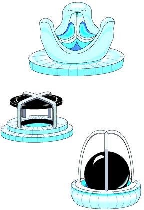
The two main types of valves are "Mechanical" and "Bioprosthetic". Mechanical valves have simply been devised from scratch, and are made of metal or similar materials. Bioprosthetic valves are fashioned from animal or human tissues.
Mechanical valves. There are several different varieties, including "bileaflet tilting valves" which are extremely popular and reliable. Tilting single disc devices are also quite popular. Older models included the "caged-ball" device and "floating discs". The body does not recognize these as "foreign", and thus there is not a fear of rejection. Mechanical valves are generally felt to have the advantage of lasting the longest time. Their main disadvantage compared to other types of valves is the need to take potent blood thinners which decreases the tendency to form clots on their surface.
Bioprosthetic valves. These use some biologic material in their composition. They are all treated, and do not carry the risk of rejection. Treated aortic valves from human cadavers, treated pig aortic valves, and valves fashioned from the pericardium (the outside lining of the heart) of cows are all utilized. These types of valves do not necessarily require that the patient take blood-thinners, but generally do not last as long.
Please note that there are pictures of various valves below.
What is the best kind of artificial valve?
It very much depends on the situation.
The need and ability to maintain long-term anticoagulation (blood thinning) is a very important factor. Several classes of people may not be good candidates to take warfarin (brand name coumadin), and therefore should receive a bioprosthetic valve. This group includes young women who wish to become pregnant. They should not take warfarin since it can frequently cause birth defects. Some people wish to continue to lead very, very active lives and taking a blood thinner would complicate the ability to do so. People who have had previous and sometimes repeated problems with bleeding (for example, frequent bleeding from ulcers) are often felt to be better served with the bioprosthetic valves which do not require anticoagulation.
On the other hand, for people who are going to need to have anticoagulation anyhow because they have the abnormal rhythm known as atrial fibrillation, using a bioprosthetic valve would have little advantage since they are going to need to take the blood thinner anyhow. They will most often receive a mechanical valve to take advantage of its longer life.
Another consideration for some people is the efficiency of the valve. That is, the replacement valve is never as good as the "real thing" in terms of how well it opens. Some artificial valves obstruct the flow of blood quite a bit, while others (mainly some of the bioprosthetic varieties) obstruct very little. There are some world-class athletes who compete despite having artificial valves, and they will have one of these "low profile" valves utilized to avoid loss of efficiency. This is generally not a consideration for most of us.
I have an artificial valve -- do I need to take blood thinners?
Many people with mechanical valves will need to take warfarin to prevent clots from forming on the artificial valve. These clots may then travel to other parts of the body. Many patients with the abnormal rhythm known as atrial fibrillation will also need to take warfarin regardless of the type of valve which is placed. Aspirin or other agents may be used instead of, or in addition to warfarin.
What is "Endocarditis"?
Endocarditis is an infection of the heart valves and parts of the inside lining of the heart muscle (known as the "endocardium"). This is an uncommon, but not rare, infection. It is often very serious. The infection may begin at the time of a dental or medical procedure in someone who has a predisposing abnormality of their heart. It may also occur in someone who has had no previous problem with their heart, and who has not had any other procedure which is associated with a risk of endocarditis.
For more about this condition, please follow the link below.To learn more about specific valvular problems, follow the links:
· Mitral Valve Prolapse
· Endocarditis
· Problems with the Aortic Valve: Aortic Stenosis and Aortic Regurgitation (Coming Soon)
· Problems with the Mitral Valve: Mitral Stenosis and Mitral Regurgitation (Coming Soon)
· Problems with the Pulmonic and Tricuspid Valves (Coming Soon)
©COPY 1997 HeartPoint Updated May 1998

![]()
| Commentary |
Food You Will Love | HeartPoint
Gallery | In The News | Health Tips | What's New
| Information Center | Home
|
This site presents material for your information, education and entertainment. We can assume no liability for inaccuracies, errors, or omissions. Above all, material on this site should not take the place of the care you receive from a personal physician. It is simply designed to help in the understanding of the heart and heart disease, and not as a diagnostic or therapeutic aid. You should seek prompt medical care for any specific health issues. Please feel free to browse the site and download material for personal and non-commercial use. You may not however distribute, modify, transmit or reuse any of these materials for public or commercial use. You should assume that all contents of the site are copyrighted. ©COPY;1997 HeartPoint